X-ray image acquisition and diagnostic software package for CR systems

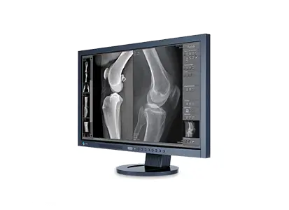
dicomPACS®DX-R  is a professional acquisition software for X-ray images generated by CR units (computed radiography with imaging plates). The software also controls the operation of X-ray generators and X-ray units manufactured by diverse companies, thus ensuring an efficient and orderly workflow. The straightforward and user-friendly GUI (graphical user interface) functions via touchscreen and mouse. dicomPACS®DX-R  produces images of outstanding quality and can be adapted to special customer needs. High-performance image processing allows organ-specific optimisation.

Everyday medical care is made easier by an array of integrated functions (e.g., a multimedia X-ray positioning guide) and an intuitive design. dicomPACS®DX-R  software can readily be integrated with existing information management systems. Furthermore, X-ray images can be evaluated using the dicomPACS®viewer module included in the acquisition software. Thus, the system functions as a fully-fledged diagnostic workstation with the option to upgrade to a PACS (Picture Archiving and Communication System).

Make dicomPACS®DX-R  the linchpin of your direct digital X-ray system - be it a new unit with generator control, a retrofit of an existing X-ray machine, or a portable suitcase solution for mobile X-ray generators.


nach oben

General benefits and features of the dicomPACS®DX-R Software

User friendliness and smooth workflow:
  • The following CR systems are supported:
    -    Carestream Point of Care (PoC 120 – 360)
    -    Carestream Vita
    -    Carestram Vita flex CR units with Kodak QC software version 2.5 or higher
    -    3DISC Imaging FireCR+ and FireCR flash
    -    3DISC Imaging FireCR dental
    -    FUJI Prima T2 (OEM version)
  • Modern graphical user interface (GUI) adaptable to almost any language 
  • Touchscreen operation – ensures a quick, efficient and orderly workflow
  • Patient data are captured via DICOM Worklist, BDT/GDT, HL7 or other protocols – data can also be captured manually
  • DICOM procedure codes are used to transfer all records relevant to an examination directly from associated information management systems (e.g., HIS/RIS)
  • Over 400 projections and a multitude of adjustment parameters are available to facilitate visualizing images of human body parts stored in the system
  • Safe and quick registration of emergency patients 
  • Scheduled examination order can be revised post factum in order to avoid frequent patient repositioning
  • Images can be appended to records even after examinations are complete
  • Additional special functions for human medicine including Chiro Tools (diagnostic tools for efficient analyses) and tools that assist with NUCCA examinations
  • User-defined macros for recurring examinations, e.g. thorax screenings 
  • Fully integrated radiographic positioning guide for all examinations in human medicine with comprehensive notes, photographs, videos and sample X-ray images
  • Wireless remote control of digital X-ray system, with display of worklist, image thumbnails and much more...
nach oben

Other advantages of dicomPACS®DX-R

The professsional dicomPACS®DX-R image processing:
  • Perfect images every time – few adjustments needed
  • Integrated software for automatic image optimisation
  • Professional image processing – adaptable to different types of examinations according to customer demands 
  • Special image processing procedures ensure optimal images over a wide range of X-ray parameter settings (e.g., dose reduction) 
  • Bones and soft tissue can be examined in one image for a better diagnosis
  • Details of bones and microstructures are very easy to recognise
  • Noise reduction
  • Black mask (automatic shutters)
  • Automatic removal of grid lines when using fixed grids
Outstandingly sophisticated image diagnosis, e.g.:
  • Integrated dicomPACS viewer for image diagnosis, processing, and storage in an SQL database, including image manipulation, export options, layout adjustments, user interface customisation and much more
  • Stepless zoom, PAN, magnification tool, ROI, crop, rotate, mirror etc.
  • Insertion of image annotations, e.g. text, arrow, ellipse
  • Measurement of distances, angles, areas and density
  • Adjustment of window/level options as well as gamma correction, sharpening filters and noise suppression 
  • Many additional functions such as Chiro Tools, calculation of Cobb's angle, pelvic obliquity measurements, automated generation of medical reports etc. 
  • Upgrade to integrated image management system (PACS) possible 
Exporting and distributing images
  • Worldwide distribution of images to medical staff and patients via dicomPACS® web server, images can be exported in JPEG, TIFF, BMP and DICOM formats
  • Images can be printed with Windows OS printers as well as laser imagers via DICOM Basic Print
  • DICOM patient CD can be created with the free WEB viewer
  • Optional external archive via web server 
  • Built-in email tool for image distribution - no external email application necessary
  • Image distribution to multiple recipients via image management systems and DICOM Store 
nach oben
  • Product brochure acquisition and diagnostic software for X-ray images
    dicomPACS®DX-R is a professional acquisition software for X-ray images from flat panel systems (DR) and CR units (computed radiography with imaging plates) by any manufacturer. In addition, the software controls X-ray generators and X-ray units of various manufacturers, providing a smooth and systematic workflow.

  • Overview of integrated components
    Integrated components: intergrated flat panels, CCD systems, X-ray generators and CR systems

  • Product overview Digital X-ray in human medicine
    Product portfolio of OR Technology: Digital Radiography and Image Management - A guide for medical practices, clinics and hospitals

nach oben

Optional add-ons

PACS MobileView

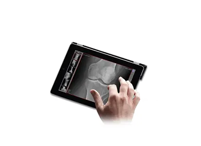
dicomPACS®MobileView - the web-based viewer for mobile devices
The web-based viewer contains all the basic functions for viewing images. The viewing can take place virtually indpendant from the browser on mobile devices, such as an iPad. dicomPACS®MobileView offers doctors and nursing staff a previously unknown, mobile freedom in the workplace inside and outside of hospitals or practices, with the radiological image material available at all times.

ORCA


Archival storage of images and document as well as communication via the internet with ORCA
ORCA
(Oehm and Rehbein Cloud Archiving) is a cloud-based platform specially designed for storing, viewing and sharing medical images and documents. ORCA offers two exciting applications: ORCA Archive and ORCA Share.


PACS Software

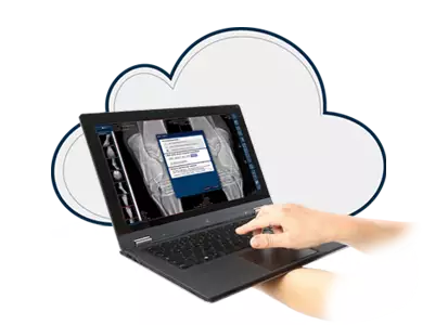
dicomPACS® – the sophisticated and high-tech image management solution  
With dicomPACS® all images generated by digital X-ray, CT, MRI and ultrasound devices, as well as diverse documents (e.g. doctor's letters, medical findings, medical history, faxes) are stored in a digital patient folder and readily accessible. 


nach oben
Subject to technical modifications