Acquisition software for static and dynamic X-ray images from flat panel or CR systems

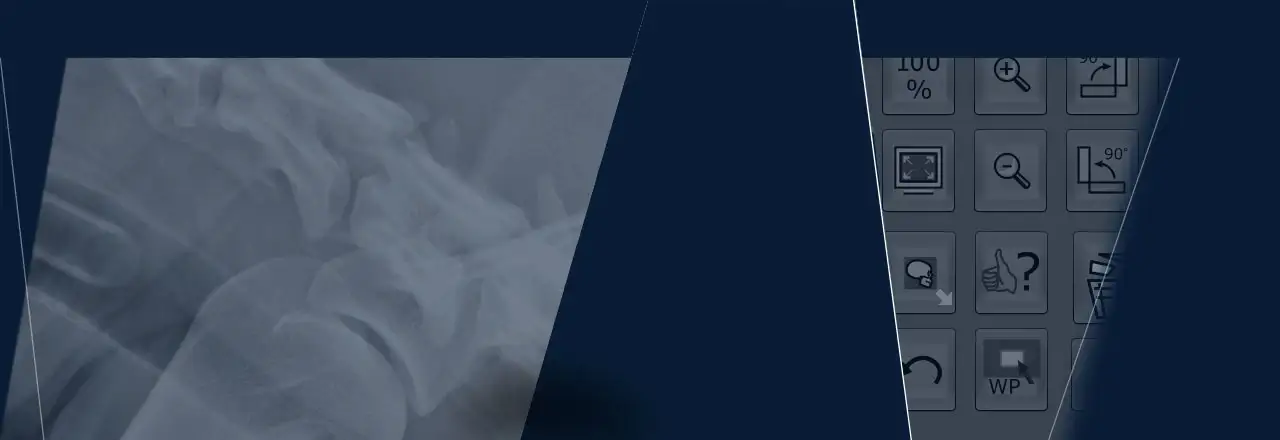
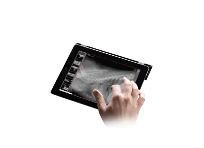
dicomPACS®DX-R is a professional acquisition software for X-ray images generated by various flat panel systems (DR) and CR units (computed radiography with imaging plates). The software also controls the operation of X-ray generators and X-ray units manufactured by diverse companies, thus ensuring an efficient and orderly workflow. The straightforward and user-friendly GUI (graphical user interface) functions via touchscreen and mouse. dicomPACS®DX-R produces images of outstanding quality and can be adapted to special customer needs. High-performance image processing allows organ-specific optimisation.

Everyday medical care is made easier by an array of integrated functions (e.g., a multimedia X-ray positioning guide) and an intuitive design. dicomPACS®DX-R software can readily be integrated with existing information management systems. Furthermore, X-ray images can be evaluated using the dicomPACS®viewer module included in the acquisition software. Thus, the system functions as a fully-fledged diagnostic workstation with the option to upgrade to a PACS (Picture Archiving and Communication System).

Make dicomPACS®DX-R the linchpin of your direct digital X-ray system - be it a new unit with generator control, a retrofit of an existing X-ray machine, or a portable suitcase solution for mobile X-ray generators.


nach oben

General benefits and features of the dicomPACS®DX-R software

User friendliness and smooth workflow:
  • Modern graphical user interface (GUI) adaptable to almost any language 
  • Touchscreen operation – ensures a quick, efficient and orderly workflow
  • Patient data are captured via DICOM Worklist, BDT/GDT, HL7 or other protocols – data can also be captured manually
  • DICOM procedure codes are used to transfer all records relevant to an examination directly from associated information management systems (e.g., HIS/RIS)
  • Over 400 projections and a multitude of adjustment parameters are available to facilitate visualizing images of animal body parts stored in the system
  • Safe and quick registration of emergency patients 
  • Scheduled examination order can be revised post factum in order to avoid frequent patient repositioning
  • Images can be appended to records even after examinations are complete
  • Special features for veterinary medicine, including a dialogue box for patient and owner data, integrated hip dysplasia measurements, special image filters, multi-generator operation for alternating between mobile and stationary X-ray systems and much more…
  • Using an algorithm based on artificial intelligence (AI), the HD measurement is inserted automatically. The survey can then be edited manually.
  • Additional special functions for veterinary medicine 
  • User-defined macros for recurring examinations, e.g. pre-purchase examination for horses
  • Fully integrated radiographic positioning guide for all examinations with comprehensive notes, photographs, videos and sample X-ray images
  • Wireless remote control of digital X-ray system, with display of worklist, image thumbnails and much more...
Flexible image acquisition:
  • Integration of various flat panel and CR systems  by different manufacturers for static and dynamic X-ray [more about dynamic X-ray]
  • Up to three flat panel detectors (Bucky, wall stand and portable) can be connected to one system
  • The generator interface of the software can be configured to control the X-ray generators and X-ray systems of diverse manufacturers.
  • The software offers parallel operation of flat panel and CR systems. The user determines which device will capture the next image. Excellent fail-safe option in case of a defect flat panel detector.
  • AEC (Automatic Exposure Control) and ARP (Anatomical Programmed Radiography) automatically set X-ray parameter values for each exposure; manual adjustments can easily be made
  • Integration of dose area product meters (DAP) – settings are saved directly to relevant image
  • Electronic X-ray log (optional)
nach oben

Other advantages of dicomPACS®DX-R

The professsional dicomPACS®DX-R image processing:
  • Perfect images every time – few adjustments needed
  • Integrated software for automatic image optimisation
  • Professional image processing – adaptable to different types of examinations according to customer demands 
  • Special image processing procedures ensure optimal images over a wide range of X-ray parameter settings (e.g., dose reduction) 
  • Bones and soft tissue can be examined in one image for a better diagnosis
  • Details of bones and microstructures are very easy to recognise
  • Noise reduction
  • Black mask (automatic shutters)
  • Automatic removal of grid lines when using fixed grids
  • ADPC (automatic dead pixel correction): Automatically eliminates dead pixels – this reduces the need to calibrate the flat panel
  • AIAA (automatic image area analysis): Automatically analyses each image for soft tissue and bone structures and applies the most suitable image processing algorithms
  • MFLA (multi frequency level analysis): Analyses each image on various frequency levels for ideal sharpness and high subtle contrast
  • ANF (automatic noise filter): Algorithm for optimal noise reduction 
  • GLI (gridless imaging): Exposures without grid. Enables the display of an image as if it had been taken with a grid [more about GLI - X-ray without grid]
  • AGLS (automatic grid line suppression): Automatically removes gridlines from flat panel images, suitable for grids from 100 LPI to 200 LPI 
  • IBC (intelligent brightness control): Automatically displays the image at the ideal level of brightness 
  • ACO (automatic contrast optimisation): Automatic contrast equalisation across the entire image –  this enables the optimal display of soft tissue and bones at the same time 
  • ABBS (automatic black border shutter): Automatically darkens all parts of an image outside the collimated area – varying degrees of transparency are available and manual adjustments are easy to make.
Outstandingly sophisticated image diagnosis, e.g.:
  • Integrated dicomPACS®vet viewer for image diagnosis, processing, and storage in an SQL database, including image manipulation, export options, layout adjustments, user interface customisation and much more
  • Stepless zoom, PAN, magnification tool, ROI, crop, rotate, mirror etc.
  • Insertion of image annotations, e.g. text, arrow, ellipse
  • Measurement of distances, angles, areas and density
  • Special functions for veterinary medicine (e.g., filters for optimal bone and soft tissue imaging, measurement tools for TPLO and TTA, MMP, distraction index and Buchanan‘s Vertebral Heart Score for dogs and cats)
  • Adjustment of window/level options as well as gamma correction, sharpening filters and noise suppression 
  • Many additional functions such as heart measurement according to Buchanan,
    various measurement tools such as MMP and TTA, integrated capturing of diagnostic reports etc.
  • AI-assisted HD measurement for dogs ( Norberg angle)
  • Upgrade to integrated image management system (PACS) possible 
Exporting and distributing images
  • Worldwide distribution of images to medical staff and patients via dicomPACS®vet web server, images can be exported in JPEG, TIFF, BMP and DICOM formats
  • Images can be printed with Windows OS printers as well as laser imagers via DICOM Basic Print (optional)
  • DICOM patient CD can be created with the free WEB viewer
  • Optional external archive via web server 
  • Built-in email tool for image distribution - no external email application necessary
  • Image distribution to multiple recipients via image management systems and DICOM Store 
nach oben

Dynamic X-Ray

Acquisition, diagnosis and archiving of image sequences [optional]

In addition to classical X-ray examinations, it is sometimes necessary to clarify special suspicious facts and unclear diagnoses further in the context of a radioscopic examination. dicomPACS®DX-R supports this procedure with the special imaging mode "Dynamic X-Ray ", in which the region to be examined is continuously X-rayed with pulsed X-rays and displayed directly on the diagnostic monitor.

Dynamic X-rays also allow the evaluation of moving structures, such as the respiration-dependent movement of the diaphragm or the beating of the heart. This examination method is also necessary for various contrast agent examinations, especially of the gastrointestinal tract, which can be excellently imaged with dicomPACS®DX-R.

dicomPACS®DX-R supports dynamic X-ray with selected X-ray detectors of different resolution and size.


Dynamic X-ray with dicom PACS ® DX-R - Easier assessment of mobile structures


Advantages of dynamic X-ray with dicomPACS®DX-R

•   Before continuous shooting, it is possible to take still images to check the exposure parameters.

•   After the recording is finished, the length of the image sequence or window level values can be adjusted, for example.

•   Various diagnostic options are available, such as playing the sequence as a single frame sequence or as a video via cine loop.

•   The recordings can be saved as uncompressed DICOM or in JPEG 2000 format.

•   The recording sequence can also be saved as a series of individual images (required for PACS systems that do not support multi-frame DICOM).


nach oben

Convenient X-ray with remote control

Your smartphone as remote control for taking and viewing images during the X-ray process

To enable you to work comfortably and quickly outside the practice or clinic rooms, we have developed the dicomPACS®DX-R remote control app.

You can control the entire process via our X-ray app on your smartphone or tablet and, for example, check, delete and repeat the exposures. Attached to your arm or the X-ray generator, it serves as a control element. You quickly gain a first impression of the images taken without having to go to the console station (tablet PC/laptop).

The graphic interface has been designed so that it can also be operated on devices with a low resolution. The dicomPACS®DX-R remote control software works independently of the operating system and the hardware of the operating device.
 

Your smartphone as remote control for x-ray examinations

Advantages of remote control

  • Enables immediate control of the X-ray images after the X-ray exposure
  • Easy installation on a mobile device 
  • No need to use the tablet PC/laptop during the entire X-ray process

Extensive functionalities

  • Uninterrupted processing of the planning list
  • Immediate review of the exposure - no running back to the X-ray case/laptop 
  • Deletion of X-ray images 
  • Repeat exposure
  • Possibility to zoom in, zoom out and rotate images without influencing dicomPACS®DX-R
  • Adjustment of brightness without influence on dicomPACS®DX-R
  • Sequence of scheduled images does not have to be followed
  • Adding photos to document the examination [included in the next version].
  • Status of the software recognisable (ready for X-ray / detector not yet ready)


nach oben

Video: Convenient X-ray with the smartphone as remote control


nach oben
  • Product brochure acquisition and diagnostic software for X-ray images
    dicomPACS®DX-R is a professional acquisition software for X-ray images from flat panel systems (DR) and CR units (computed radiography with imaging plates) by any manufacturer. In addition, the software controls X-ray generators and X-ray units of various manufacturers, providing a smooth and systematic workflow.

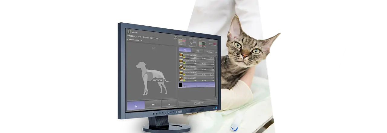
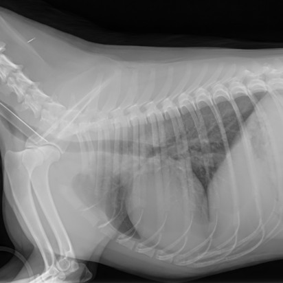
  • Product overview veterinary medicine
    Digital X-ray imaging - guide for vet clinic and practice

nach oben

Optional add-ons

PACS MobileView vet

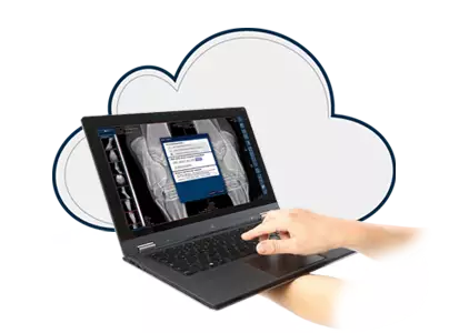
dicomPACS®MobileView - the web-based viewer for mobile devices
The web-based viewer contains all the basic functions for viewing images. The viewing can take place virtually indpendant from the browser on mobile devices, such as an iPad. dicomPACS®MobileView offers vets staff a previously unknown, mobile freedom in the workplace inside and outside of equine clinic or veterinary practice, with the radiological image material available at all times.

ORCA


Archival storage of images and document as well as communication via the internet with ORCA
ORCA
(Or Technology Cloud Archiving) is a cloud-based platform specially designed for storing, viewing and sharing medical images and documents. ORCA offers two exciting applications: ORCA Archive and ORCA Share.


nach oben

GLI (gridless imaging): X-ray imaging without grid

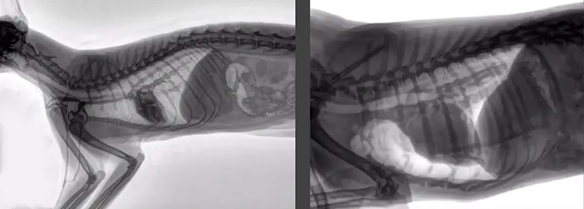
Grids are required for X-raying large body parts in order to focus the radiation and reduce scatter, thus improving the contrast and brilliance of X-ray images. The virtual scatter reduction GLI works like a grid and can be used instead of a physical grid for all body regions, including thorax, abdomen, skull, spine, pelvis and upper and lower extremities.

Move the blue slider for a comparison:
left: without grid / right: with GLI

Acquisition and diagnostics software for veterinarians with GLI (gridless imaging)
GLI (gridless imaging) emulated raster properties with flexible, customisable virtual raster


Improved image contrast and brilliant images through GLI

The grid properties emulated virtually with GLI (gridless imaging) can be flexibly adapted to act like a physical grid. As a result, intelligent image processing with GLI significantly improves the contrast and brilliance of X-ray images without a grid while simultaneously reducing the applied dose. GLI eliminates problems such as uneven brightness or cropped image areas caused by improper alignment or focusing of a physical grid.

The use of GLI is very convenient, as no antiscatter grid needs to be positioned for the X-raying process. GLI is a considerable advantage for the x-ray of non-sedated animals, as the exposure times can be shortened significantly due to the dose reduction, thus minimizing the risk of motion blur in the x-ray images.


nach oben
Subject to technical modifications