X-ray software wanted

OEM partnerships provide numerous benefits to manufacturers who are interested in combing their X-ray systems with our dicomPACS®DX-R acquisition software under a chosen brand name. Taking advantage of an existing X-ray software reduces development time and costs.

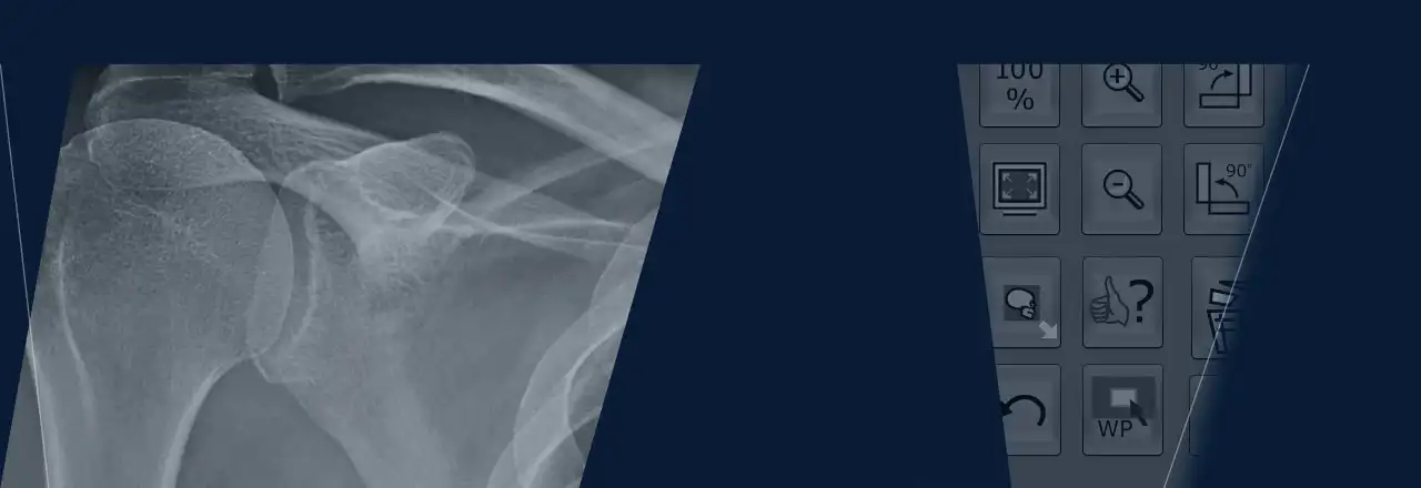
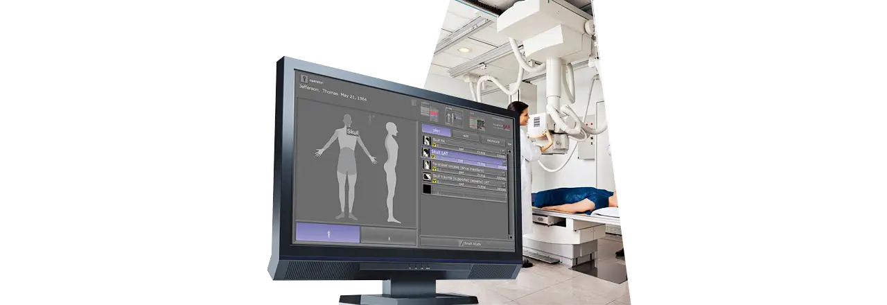
dicomPACS®DX-R is THE software for the complete integration of X-ray generators, stands, detectors, CR systems, image processing, file management, patient administration and PACS (including cloud computing). dicomPACS®DX-R is a professional acquisition software for static and dynamic X-ray images generated by various X-ray detector systems (DR) and CR units (imaging plates reader). The software also controls the operation of X-ray generators and X-ray units manufactured by diverse companies, thus ensuring an efficient and orderly workflow. The user-friendly and straightforward visual interface functions via touchscreen and mouse.

Daily work routines are made easier by an intuitive design and an array of integrated functions. This includes a multimedia X-ray positioning guide as well as special aids for human medicine, e.g., Chiro Tools (diagnostic tools for efficient analyses) and a NUCCA tool set as well as as the possibility of diagnosis supported by an automatic, AI-assisted thorax screening (by a qualified third-party provider) is also included in the software (optional).

dicomPACS®DX-R software can readily be integrated with existing information management systems. Furthermore, X-ray images can be evaluated using the dicomPACS® viewer module included in the acquisition software. Thus, the system functions as a fully-fledged diagnostic workstation with the option to upgrade to a PACS (Picture Archiving and Communication System).


nach oben

Benefits of the dicomPACS®DX-R software (CE, FDA 510[k] and Health Canada certified):

User friendliness and smooth workflow:
  • Modern graphical user interface (GUI) adaptable to almost any language 
  • Touchscreen operation – to ensure quick and efficient work and a smooth workflow
  • Capture of patient data via DICOM Worklist, BDT/GDT, HL7 or other protocols – data may also be captured manually
  • Use of DICOM Procedure Codes for the transfer of all relevant examination data directly from the connected patient management system (HIS/RIS)
  • Freely configurable body parts with more than 400 projections and numerous possible adjustments in human and veterinary medicine already included
  • Safe and fast registration of emergency patients
  • Allows the user to switch between examinations of a patient, for instance to avoid having to re-position the patient frequently
  • Allows the user to subsequently add images to an examination, even after that examination has already been completed
  • Special tools for veterinary medicine, such as an extra dialog box for patient and owner data, integrated hip dysplasia measuring, special image filters, multi generator operation for alternating between mobile and stationary X-ray systems and much more…
  • Entry of recurring examination procedures as macros, e.g. thorax screenings or pre-purchase examination for horses
  • Fully integrated radiographic positioning guide for each examination in human and veterinary medicine incl. comprehensive notes, photos, videos and correct X-ray images
  • Option to control a digital X-ray system via wireless remote incl. display of the worklist, preview of the image taken for checking and much more
Flexible image acquisition:
  • Integration of various flat panel and CR systems  by different manufacturers for static and dynamic X-ray [more about dynamic X-ray]
  • Option to connect up to 3 flat panels (bucky, wall stand and mobile) to one system
  • The configurable generator interface enables the user to control X-ray generators or X-ray systems by different manufacturers, delivering the generator settings directly from the software
  • Option for the parallel operation of a flat panel and a CR system included in the standard package. The user has the choice to take the next image with either the flat panel or the integrated CR system. This flexibility also provides an excellent emergency concept in case of a defect flat panel.
  • AEC (Automatic Exposure Control) and APR (Anatomical Programmed Radiography) allow the user to automatically adjust all X-ray options for each projection with an option to subsequently edit the image manually
  • Integration of dose area product meters (DAP) – the readings are saved directly to the relevant image
  • Electronic X-ray log
nach oben

Dynamic X-Ray

Acquisition, diagnosis and archiving of image sequences [optional]

In addition to classical X-ray examinations, it is sometimes necessary to clarify special suspicious facts and unclear diagnoses further in the context of a radioscopic examination. dicomPACS®DX-R supports this procedure with the special imaging mode "Dynamic X-Ray ", in which the region to be examined is continuously X-rayed with pulsed X-rays and displayed directly on the diagnostic monitor.

Dynamic X-rays also allow the evaluation of moving structures, such as the respiration-dependent movement of the diaphragm or the beating of the heart. This examination method is also necessary for various contrast agent examinations, especially of the gastrointestinal tract, which can be excellently imaged with dicomPACS®DX-R.

dicomPACS®DX-R supports dynamic X-ray with selected X-ray detectors of different resolution and size.


Dynamic X-ray the special acquisition mode supports the assessment of moving structures


Advantages of dynamic X-ray with dicomPACS®DX-R

•   Before continuous shooting, it is possible to take still images to check the exposure parameters.

•   After the recording is finished, the length of the image sequence or window level values can be adjusted, for example.

•   Various diagnostic options are available, such as playing the sequence as a single frame sequence or as a video via cine loop.

•   The recordings can be saved as uncompressed DICOM or in JPEG 2000 format.

•   The recording sequence can also be saved as a series of individual images (required for PACS systems that do not support multi-frame DICOM).


nach oben

Convenient X-ray with remote control

Your smartphone as remote control for planning and viewing images during the X-ray process

To enable you to work comfortably and quickly outside the practice or clinic rooms, we have developed the dicomPACS®DX-R remote control app.

You can control the entire process via our X-ray app on your smartphone or tablet and, for example, check or delete. Attached to your arm or the X-ray generator, it serves as a control element. You quickly gain a first impression of the images taken without having to go to the console station (tablet PC/laptop).

The graphic interface has been designed so that it can also be operated on devices with a low resolution. The dicomPACS®DX-R remote control software works independently of the operating system and the hardware of the operating device.

Ihr Smartphone als Fernbedienung für die Aufnahme und Betrachtung von Bildern während des Röntgenvorganges

Advantages of remote control

  • Enables immediate control of the X-ray images after the X-ray exposure
  • Easy installation on a mobile device 
  • No need to use the tablet PC/laptop during the entire X-ray process

Extensive functionalities

  • Uninterrupted processing of the planning list
  • Immediate review of the exposure - no running back to the X-ray case/laptop 
  • Deletion of X-ray images 
  • Repeat planning
  • Possibility to zoom in, zoom out and rotate images without influencing dicomPACS®DX-R
  • Adjustment of brightness without influence on dicomPACS®DX-R
  • Sequence of scheduled images does not have to be followed
  • Adding photos to document the examination [included in the next version].
  • Status of the software recognisable (ready for X-ray / detector not yet ready)


nach oben

Other advantages of dicomPACS®DX-R

The professsional dicomPACS®DX-R image processing:
  • Perfect images every time – few adjustments needed
  • Integrated software for automatic image optimisation
  • Professional image processing – adaptable to different types of examinations according to customer demands 
  • Special image processing procedures ensure optimal images over a wide range of X-ray parameter settings (e.g., dose reduction) 
  • Bones and soft tissue can be examined in one image for a better diagnosis
  • Details of bones and microstructures are very easy to recognise
  • Noise reduction
  • Black mask (automatic shutters)
  • Automatic removal of grid lines when using fixed grids
  • ADPC (automatic dead pixel correction): Automatically eliminates dead pixels – this reduces the need to calibrate the flat panel
  • AIAA (automatic image area analysis): Automatically analyses each image for soft tissue and bone structures and applies the most suitable image processing algorithms
  • MFLA (multi frequency level analysis): Analyses each image on various frequency levels for ideal sharpness and high subtle contrast
  • ANF (automatic noise filter): Algorithm for optimal noise reduction 
  • GLI (gridless imaging): Exposures without grid. Enables the display of an image as if it had been taken with a grid – useful for supine chest exposures (bedside)
  • AGLS (automatic grid line suppression): Automatically removes gridlines from flat panel images, suitable for grids from 100 LPI to 200 LPI 
  • IBC (intelligent brightness control): Automatically displays the image at the ideal level of brightness 
  • ACO (automatic contrast optimisation): Automatic contrast equalisation across the entire image –  this enables the optimal display of soft tissue and bones at the same time 
  • ABBS (automatic black border shutter): Automatically darkens all parts of an image outside the collimated area – varying degrees of transparency are available and manual adjustments are easy to make.
Outstandingly sophisticated image diagnosis, e.g.:
  • Integrated dicomPACS viewer for image diagnosis, processing, and storage in an SQL database, including image manipulation, export options, layout adjustments, user interface customisation and much more
  • Stepless zoom, PAN, magnification tool, ROI, crop, rotate, mirror etc.
  • Insertion of image annotations, e.g. text, arrow, ellipse
  • Measurement of distances, angles, areas and density
  • Extended acquisition functionality for use in mammography*: Specialised organ tree, mammography Hanging Protocols, documentation of additional examination parameters such as filter, thickness, angle, implant and many more
  • Adjustment of window/level options as well as gamma correction, sharpening filters and noise suppression 
  • Many additional functions such as Chiro Tools, calculation of Cobb's angle, pelvic obliquity measurements, automated generation of medical reportsas well as tas the possibility of diagnosis supported by an automatic, AI-assisted thorax screening (by a qualified third-party provider) etc. 
  • Upgrade to integrated image management system (PACS) possible 
  • Extended acquisition functionality for use in mammography*: Specialised organ tree, Mammography Hanging Protocols, Documentation of additional examination parameters such as filter, thickness, angle, implant and many more

    *Pending CE and FDA conformity approval for the module
Exporting and distributing images
  • Worldwide distribution of images to medical staff and patients via dicomPACS® web server, images can be exported in JPEG, TIFF, BMP and DICOM formats
  • Images can be printed with Windows OS printers as well as laser imagers via DICOM Basic Print
  • DICOM patient CD can be created with the free WEB viewer
  • Optional external archive via web server 
  • Built-in email tool for image distribution - no external email application necessary
  • Image distribution to multiple recipients via image management systems and DICOM Store 
nach oben
  • Product brochure dicomPACS®DX-R Acquisitions and diagnostic software
    Acquisition and diagnostic software for X-ray images from DR flat panels or CR systems in human and veeterinary medicine

  • Overview of integrated components
    Integrated components: integrated flat panels, CCD systems, X-ray generators and CR systems

  • Product overview Digital X-ray in human medicine
    Product portfolio of OR Technology: Digital Radiography and Image Management - A guide for medical practices, clinics and hospitals

nach oben

GLI (gridless imaging): X-ray imaging without grid

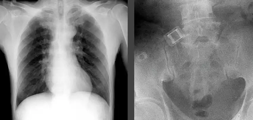
Grids are required for X-raying large body parts in order to focus the radiation and reduce scatter, thus improving the contrast and brilliance of X-ray images. The virtual scatter reduction GLI works like a grid and can be used instead of a physical grid for all body regions, including thorax, abdomen, skull, spine, pelvis and upper and lower extremities.

Move the blue slider for a comparison:
left: without grid / right: with GLI

GLI gridless imaging digital X-ray without grid


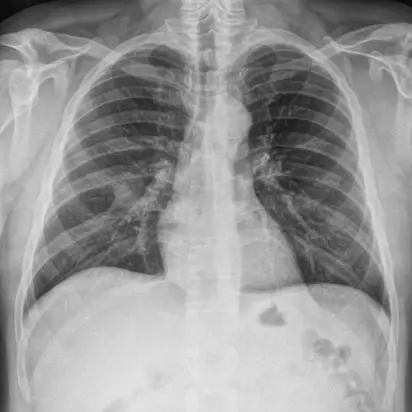
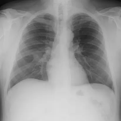
Improved image contrast and brilliant images through GLI

The grid properties emulated virtually with GLI (gridless imaging) can be flexibly adapted to act like a physical grid. As a result, intelligent image processing with GLI significantly improves the contrast and brilliance of X-ray images without a grid while simultaneously reducing the applied dose. GLI eliminates problems such as uneven brightness or cropped image areas caused by improper alignment or focusing of a physical grid.

GLI can be used in many clinical scenarios - at the hospital bedside, in the operating / emergency room - in which the positioning of a physical grid is a challenge or an optimal grid is not available. In these often mobile applications, GLI improves the workflow without antiscatter grid.

nach oben
Subject to technical modifications